
Key Takeaways
- RI-MUHC researchers created spontaneously beating 3D cardiac tissue from blood cells of a patient with dilated cardiomyopathy.
- The patient-derived tissue showed abnormal, irregular beating patterns and other features consistent with the patient’s disease.
- The work is a proof of concept that could support patient-specific therapy research and preclinical drug development.
How Do You Turn Blood Into Beating Heart Tissue?
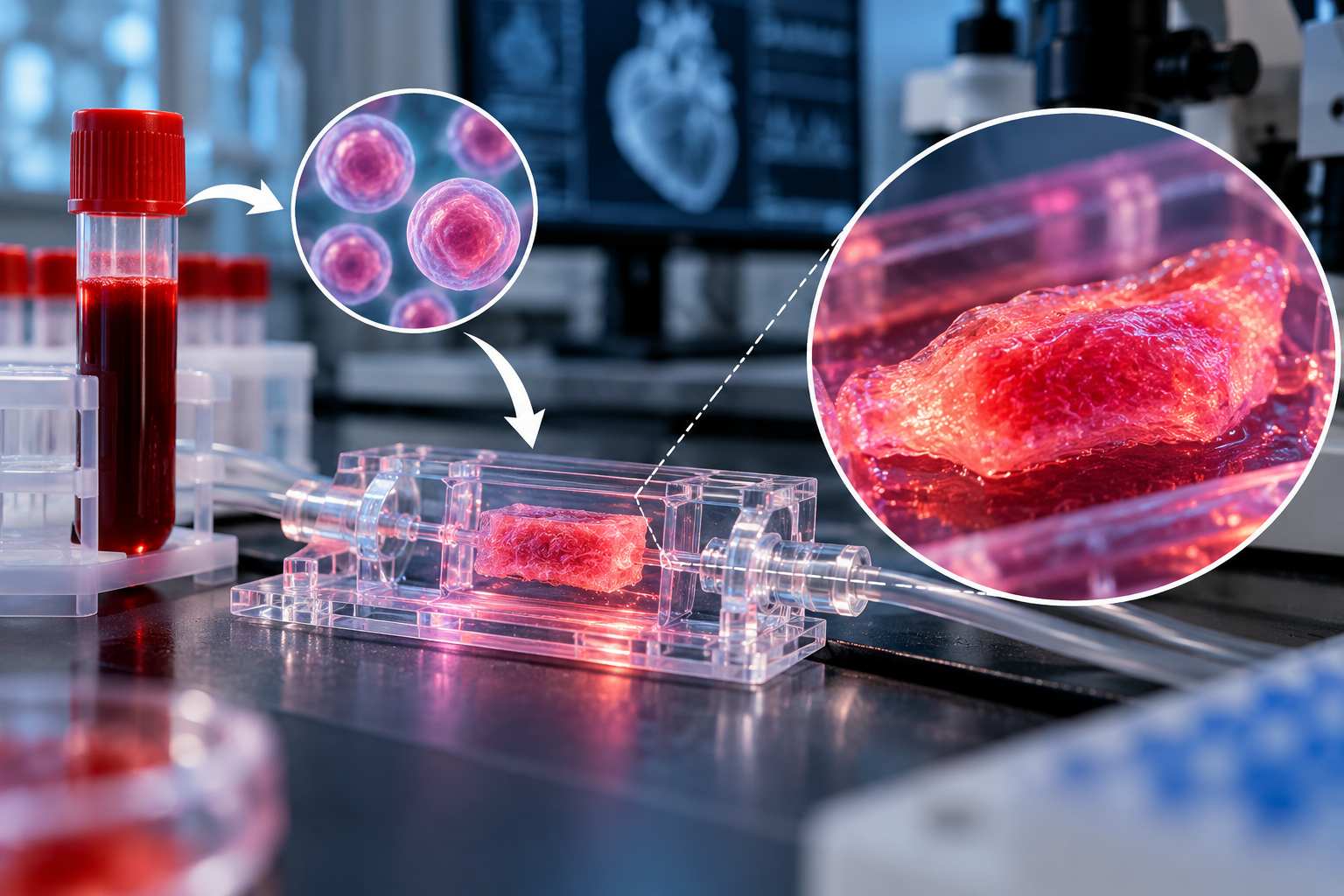
The process starts with blood cells from a patient.
Researchers reprogrammed those cells into induced pluripotent stem cells, or iPSCs.
These are reprogrammed cells that can develop into many different cell types in the human body.
The scientists then transformed the patient-specific stem cells into cardiomyocytes — the muscle cells responsible for the heart's contractions.
Those cells were used to create three-dimensional cardiac tissue on a chip.
The result was engineered tissue capable of beating and contracting spontaneously.
Essentially, researchers were able to take cells originating from a patient's blood and use them to recreate certain characteristics of that person's heart disease outside the body.
Why This Particular Heart Disease?
The patient had dilated cardiomyopathy, or DCM.
In DCM, the heart becomes enlarged and weakened, making it more difficult to pump blood effectively. The condition can lead to heart failure and abnormal heart rhythms and is a leading cause of heart transplantation.
Studying diseases like DCM is challenging.
A dish of cells isn't a human heart, and an animal heart isn't identical to one either. Researchers therefore need better laboratory models that can reproduce important features of human heart disease.
A patient-derived heart-on-a-chip offers another possibility: create living cardiac tissue carrying characteristics of the patient's disease and watch how it behaves in the laboratory.
The “Mini-Heart” Behaved Differently
The researchers didn't simply create the tissue and watch it beat.
They compared it with engineered cardiac tissue created using cells from a healthy participant.
The differences were measurable.
Researchers observed differences in the tissues' structure, contractions, beating patterns, calcium activity and molecular characteristics.
Most strikingly, the cardiac tissue derived from the DCM patient showed abnormal and irregular beating patterns consistent with important features of the disease.
Researchers also exposed the engineered tissues to norepinephrine, which can increase heart rate and the force of contraction.
The tissues responded to the stimulation.
That gives researchers another way to examine how patient-derived cardiac tissue behaves under different conditions.
Tiny Beads Help Scientists Watch Every Beat
One particularly clever part of the experiment involved something surprisingly simple:
tiny fluorescent beads.
Researchers embedded the beads directly into the engineered tissue.
As the tissue contracted, scientists could track how the beads moved, allowing them to map contractions across different regions of the tissue.
Instead of simply seeing whether the tissue was beating, researchers could examine how those contractions occurred across the engineered cardiac tissue.
Could Your Future Medicine Be Tested on Your Own Cells?
That is where this research becomes especially interesting — but also where it's important not to get ahead of the science.
The researchers describe the work as a proof of concept.
It does not mean doctors can currently take every heart patient's blood, create a miniature heart and use it to select their treatment.
But the research points toward a potentially powerful future application.
Scientists could eventually create patient-specific cardiac models and investigate how those tissues respond to different therapies.
The platform could also help with preclinical drug development by giving researchers another way to study potential treatments before they reach patients.
And because the system uses human-derived cardiac tissue, it could complement existing laboratory and animal research and potentially reduce reliance on animal models for some experiments.
The Next Challenge Is Much Bigger
One patient-derived disease model is an intriguing demonstration.
But it isn't evidence that the technology will work across the enormous biological diversity of people living with heart disease.
That's precisely what researchers want to investigate next.
The team plans to expand the platform to samples from a much larger number of patients, including people with different genetic backgrounds and different forms of dilated cardiomyopathy.
If researchers can reliably reproduce individual variations in heart disease, the technology could move closer to a much bigger goal:
studying a patient's disease using heart tissue created from that patient's own cells.
For now, this remains laboratory research.
But turning a blood sample into beating cardiac tissue that reproduces important features of a patient's disease offers a fascinating glimpse at where personalized heart research could be heading.