
Key Takeaways
- Aggressive breast cancer cells invaded engineered liver tissue more readily than engineered cardiac tissue.
- The MInD platform connects multiple engineered tissues under fluid flow to study cancer-cell invasion.
- The study does not create a cancer treatment, but it may help identify mechanisms that limit metastasis.
Why Cancer Spreading Matters So Much
Cancer becomes particularly dangerous when malignant cells escape the original tumour, travel through the body and establish new tumours elsewhere.
This process is known as metastasis.
Different cancers have different preferred destinations. Breast cancer, for example, can metastasize to organs and tissues including the bones, liver, lungs and brain.
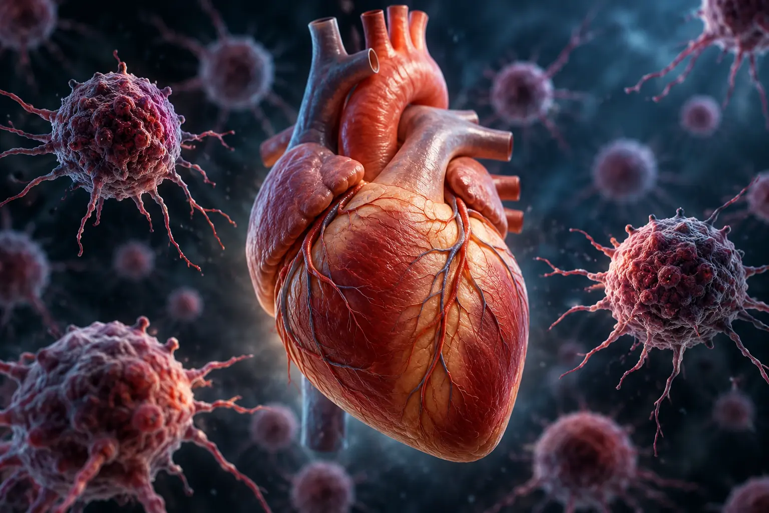
The ventricular myocardium — the muscular tissue that makes up much of the heart's pumping chambers — is one of the least common sites of metastasis.
That raises a fascinating biological question.
If cancer cells can travel through the body and reach many different organs, what makes some tissues much easier to invade than others?
Researchers at the University of Toronto's Institute of Biomedical Engineering wanted a better way to investigate that question.
Scientists Built Multiple Tiny Tissue Environments
Studying metastasis is difficult.
Traditional cell cultures can be useful, but they cannot fully recreate the complicated environment inside human organs. Animal models provide much greater biological complexity, but biological differences between animals and humans can limit how closely they reproduce human cancer metastasis.
The Toronto researchers developed another approach.
Their system is called the Multi-organ Invasion Device, or MInD.
It is a multi-organ-on-a-chip platform that allows different engineered tissues to exist within the same laboratory system while interacting under dynamic fluid flow.
Think of it as a highly simplified miniature environment where researchers can observe cancer cells interacting with different types of tissue.
Instead of simply watching cancer cells grow in a conventional dish, scientists can compare how those cells behave when confronted with environments representing different organs.
Heart vs. Liver Produced a Striking Difference
The researchers tested highly aggressive breast cancer cells against two very different engineered tissue environments.
One represented the liver.
The other represented cardiac tissue.
The liver is a common site of breast cancer metastasis, making it a useful comparison with the heart.
Cancer cells readily migrated toward and invaded the engineered liver tissue.
When those same cells encountered cardiac tissue, however, researchers found that invasion was significantly suppressed compared with the liver environment.
The difference became even more interesting when cancer cells, liver tissue and cardiac tissue were placed together in the same device.
The cancer cells preferentially migrated toward the liver compartment.
Researchers also found that the presence of cardiac tissue reduced overall invasion.
In other words, the cardiac tissue did not simply appear to be a less favourable destination. Its presence was also associated with reduced cancer-cell invasion across the experimental system.
What Could the Heart Be Doing?
This is where the research becomes particularly interesting.
The team used cytokine profiling and RNA sequencing to investigate what was happening inside the different tissue environments.
They found that cardiac co-culture was associated with suppression of pathways related to cancer-cell metastasis and invasion while also inducing immune activation.
Researchers also observed changes involving immune-related signalling and gene programs that could provide clues about why cardiac tissue is comparatively resistant to metastatic invasion.
There is an important distinction, however.
Scientists have not discovered a cancer shield that can simply be transferred from the heart to another organ.
Instead, the study gives researchers a new experimental platform for investigating the biological mechanisms that may contribute to the heart's unusual resistance.
Identifying exactly which mechanisms matter most is the next challenge.
Why the Technology Could Matter Beyond This Study
The MInD platform itself may be nearly as interesting as the initial cancer findings.
Cancer metastasis is not determined only by the characteristics of a tumour cell. The surrounding environment can influence whether cancer cells survive, migrate, invade tissue and eventually establish another tumour.
The researchers' platform connects different tissue compartments using specially designed porous structures called PermeoTubes.
These structures allow cancer cells to move between compartments while the system operates under fluid flow, giving researchers a controlled way to observe parts of the metastatic process.
By placing several engineered tissues within one connected system, scientists can investigate how different tissue environments influence cancer-cell behaviour.
The technology could eventually allow researchers to compare additional organs, investigate molecular signals involved in metastasis and test potential therapies under more complex laboratory conditions.
Organ-on-a-chip technology more broadly is being developed as a way to reproduce selected aspects of human physiology in the laboratory, potentially providing researchers with additional tools for disease modelling and drug development.
Could the Heart Teach Scientists How to Slow Cancer's Spread?
That remains an open question.
This research does not establish a new cancer treatment, and experiments involving engineered tissues cannot reproduce everything that happens inside a living human body.
But it gives scientists something valuable: a controlled way to investigate an unusual biological phenomenon.
Why can aggressive cancer cells successfully invade one tissue while struggling to invade another?
The researchers found another intriguing clue outside the main multi-organ experiment. In separate laboratory tests, media conditioned by cardiac tissue reduced proliferation in breast cancer, lung carcinoma and colon adenocarcinoma cells.
The researchers suggest cardiac tissue may release bioactive molecules — potentially including cytokines, extracellular vesicles or extracellular-matrix proteins — that contribute to its resistance to cancer invasion.
Exactly which factors are responsible, and whether any of them could eventually have therapeutic value, remains to be determined.
That is what makes the discovery interesting rather than conclusive.
Cancer cells have an extraordinary ability to spread through the human body.
Yet some tissue environments appear much harder for them to conquer than others.
If scientists can understand what makes the heart one of those environments, it could provide another piece of the puzzle surrounding one of cancer's most dangerous abilities.