
Key Takeaways
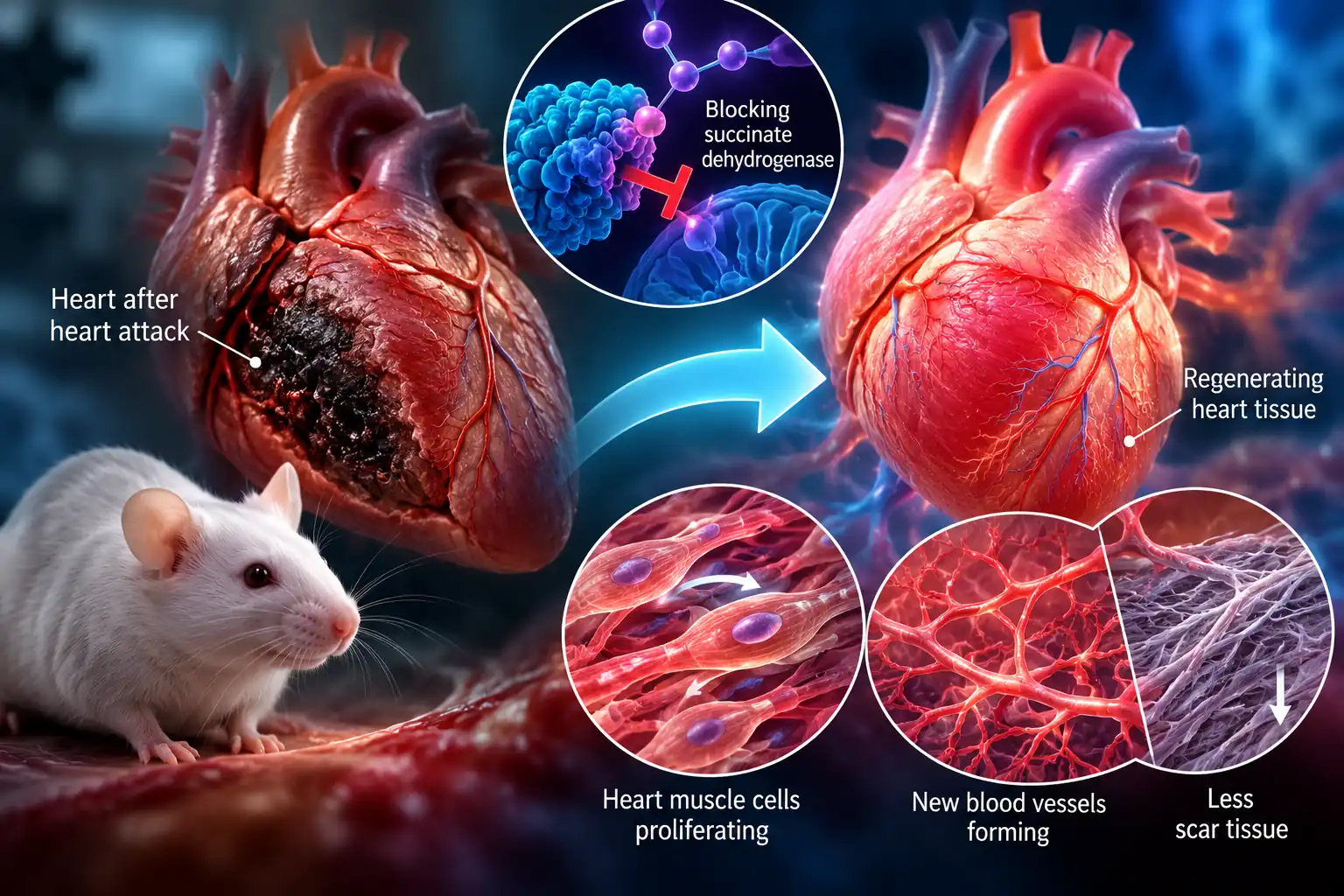
- Temporarily inhibiting succinate dehydrogenase helped damaged mouse hearts show signs of regeneration.
- Heart muscle-cell proliferation alone did not significantly improve function; fibroblast changes were also crucial.
- The findings are limited to mice and are not yet a treatment for people after heart attacks.
The adult heart is remarkably good at keeping us alive. What it is not very good at is repairing itself after a heart attack.
Scientists at Sanford Burnham Prebys Medical Discovery Institute and collaborating U.S. institutions have now shown in mice that some of the heart's regenerative machinery may not disappear entirely with age — it may become dormant.
In a peer-reviewed study published September 28, 2026, in Nature Cardiovascular Research, researchers found that temporarily changing how heart cells produce energy helped push damaged mouse hearts toward a more regenerative state.
Flipping the Heart Back Into "Repair Mode"
The researchers focused on an enzyme called succinate dehydrogenase, which plays a role in cellular energy production.
They used malonate to temporarily inhibit the enzyme, effectively changing the metabolic state of heart tissue.
The result was striking: researchers observed heart muscle cells proliferating again, new blood vessels forming and less scar tissue developing after heart attacks in mice.
The treatment appeared to recreate some features normally found in newborn hearts.
That's important because newborn mice can regenerate damaged heart tissue for a short period after birth. Adults largely lose that ability.
It Wasn't Just About Growing New Heart Cells
One of the study's more interesting discoveries came when researchers examined different types of heart cells separately.
Blocking succinate dehydrogenase specifically in heart muscle cells caused a temporary increase in their proliferation — but that alone wasn't enough to significantly improve heart function after a heart attack.
Another type of cell turned out to be crucial: cardiac fibroblasts, which are involved in forming scar tissue.
When researchers disrupted the same metabolic pathway in fibroblasts, scar-forming activity declined and cardiac function improved.
In other words, repairing the heart may require more than simply persuading muscle cells to multiply. The surrounding cellular environment also needs to change.
The Treatment Changed Which Genes Cells Could Use
The researchers found another layer to the effect.
After metabolic reprogramming, regions of DNA associated with regeneration and cell division became more accessible in heart muscle cells. At the same time, gene programs associated with fibrosis and scarring were suppressed in fibroblasts.
Senior author Ahmed Mahmoud, PhD, described the effect as coordinating regenerative responses across multiple cell types rather than activating one isolated repair pathway.
What This Does — and Doesn't — Mean for Humans
This is not yet a treatment for people who have suffered heart attacks.
The experiments were performed in mice, and therapies that produce promising results in animal models can fail to translate safely or effectively to humans.
But the study provides an intriguing clue: the adult mammalian heart's regenerative machinery may be capable of being partially reactivated by changing the metabolic state of its cells.
The researchers say their long-term objective is to move the concept toward clinical testing.
For now, the discovery raises a fascinating possibility. Instead of building an entirely new repair mechanism for the damaged heart, future therapies might one day find a way to wake up machinery that was already there.
Credits
Research: Ahmed Mahmoud, PhD; Yi Fan, PhD; Dakota Nuttall and colleagues at Sanford Burnham Prebys and collaborating U.S. institutions.
Institution: Sanford Burnham Prebys Medical Discovery Institute.
Study: A metabolic–epigenetic switch governs multicellular cardiac repair following succinate dehydrogenase inhibition.
Journal: Nature Cardiovascular Research, published September 28, 2026.
DOI: 10.1038/s44161-026-00881-9.
Source/Research Credit: Sanford Burnham Prebys / Nature Cardiovascular Research.