
Key Takeaways
- Researchers identified 31 neuronal subpopulations in the human striatum, including nine types of medium spiny neurons.
- Higher MSH2 and MSH3 expression in dorsal striatal neurons may help explain vulnerability in Huntington's disease.
- Human and mouse neurons differed in OPRM1 expression, which may limit how closely some mouse opioid studies translate to humans.
Deep inside your brain is a region that helps decide whether you move, stop, chase a reward or turn an action into a habit.
It's called the striatum.
And scientists have just created one of their most detailed maps of it yet.
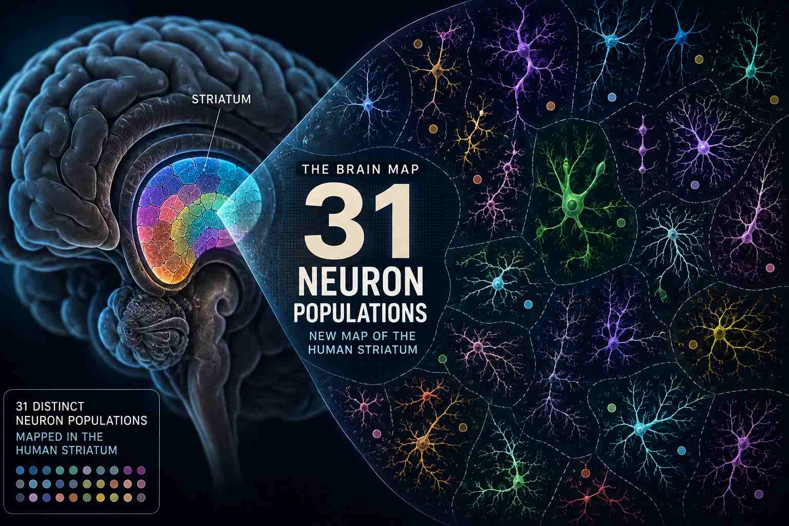
Researchers identified 31 neuronal subpopulations in the striatum — revealing differences that could help scientists better understand conditions including Huntington's disease, substance use disorder and schizophrenia.
But one of the most interesting discoveries came when researchers compared humans with mice.
Some important parts of this brain map don't match.
Quick Answer
Researchers led by scientists at MIT analyzed human brain tissue using techniques that allowed them to examine which genes were active in individual cells and where those cells were located.
They identified 31 neuronal subpopulations, including nine types of medium spiny neurons, the dominant kind of neuron in the striatum.
The study, published September 1 in Cell, also uncovered clues about why certain neurons may be particularly vulnerable in Huntington's disease — and why mice may not perfectly reproduce some aspects of the human brain's response to opioids.
Think of it less as scientists discovering a treatment and more as drawing a much better map of the territory where future treatments will have to work.
Why Map This Part of the Brain?
The striatum sits deep inside the brain, but it has an outsized job.
It helps process movement, decision-making, rewards and habits.
Scientists already knew that neurons within the striatum weren't all identical. The problem was figuring out exactly how they differed and how those differences related to disease.
So the researchers went cell by cell.
Using donated postmortem human brain tissue from brain banks in the United States and Canada, they examined patterns of gene activity and mapped where different neurons were located.
Instead of seeing one large population of essentially similar neurons, they identified 31 distinct neuronal subpopulations.
That included nine varieties of medium spiny neurons — inhibitory neurons that respond to dopamine and make up much of the striatum.
And that's where the map started becoming especially interesting.
Some Neurons May Be More Vulnerable to Huntington's Disease
Huntington's disease is caused by an inherited mutation involving repeated sections of DNA known as CAG repeats in the huntingtin gene.
Over time, the number of those repeats can expand in certain cells.
Researchers found that medium spiny neurons in the upper, or dorsal, portion of the striatum expressed higher levels of two genes called MSH2 and MSH3.
Those genes are involved in DNA mismatch repair and have been linked to the expansion of CAG repeats in the huntingtin gene.
As those repeats accumulate, the mutated huntingtin protein can become increasingly harmful to cells.
That offers a potential clue to a longstanding question:
Why are some neurons hit harder by Huntington's disease than others?
The researchers found something else intriguing.
A rare population of medium spiny neurons forming island-like structures in the ventral striatum appeared more resistant to CAG-repeat accumulation.
Understanding what makes those cells different could eventually help researchers investigate whether other vulnerable neurons can be made more resilient.
But that's a research direction — not a treatment discovered by this study.
Then the Researchers Looked at Mice
Mice are enormously important to neuroscience.
But a mouse brain isn't simply a miniature human brain.
When researchers compared their human map with mouse tissue, they found notable differences, especially in the expression of genes associated with drug responses and substance use disorders.
One stood out.
A gene called OPRM1, which encodes the mu-opioid receptor, was highly expressed in a particular human neuron population known as D1 outliers.
The corresponding mouse neurons didn't show the same pattern.
Why does that matter?
The mu-opioid receptor is central to how opioids affect the body and brain.
If an important receptor is distributed differently in humans and mice, standard mouse models may not perfectly reproduce the human biology researchers are trying to understand.
That doesn't make mouse studies useless.
Far from it.
It means scientists may need to be much more precise about which parts of mouse biology translate well to humans — and which don't.
Researchers even suggested that some mouse models could potentially be engineered to express the receptor in a more human-like way, which could make them more useful for studying certain questions involving opioid responses.
The Bigger Story Isn't “31”
The headline number is fascinating.
31 neuronal subpopulations.
But the more important discovery may be what those differences reveal.
Two neurons can live in the same brain region and still activate different genes, carry different drug-related molecular signatures and show different vulnerability to disease.
Scientists aren't simply mapping where things happen in the brain anymore.
They're increasingly mapping which individual kinds of cells are involved.
And for diseases that affect some neurons while mysteriously sparing others, that distinction could become extremely important.
TwikUp Insight
Imagine trying to repair a city while your map labels an entire neighbourhood simply as “buildings.”
A hospital, apartment tower, power station and school would all look identical.
That's roughly the problem increasingly detailed brain atlases are trying to solve.
This new map doesn't cure Huntington's disease, addiction, schizophrenia or depression.
Instead, it gives researchers something more fundamental:
a clearer idea of which cells they should actually be studying.
And sometimes, before science can figure out how to fix something, it first needs a better map.
Health & Science Disclaimer: This article is for general informational and educational purposes only. It does not provide medical advice, diagnosis or treatment. The research described here is primarily a cellular and molecular brain-mapping study and does not establish a new treatment for Huntington's disease, substance use disorder or other neurological or psychiatric conditions.